Pictorial key to common surface dwelling species of Collembola from the Netherlands
|
Checklist of the Collembola: Pictorial key to common surface dwelling species of Collembola from the Netherlands |
This key is still under construction. Note that missing figures will be provided as soon as possible. Currently, the key is in the feasibility study phase to find out how to integrate in the checklist in a modular way a key that has been generated with DELTA.
Frans Janssens,
Department of Biology, University of Antwerp, Antwerp, B-2020, Belgium
Ab H. Baas
European Invertebrate Survey - Nederland, Postbus 9517, 2300 RA Leiden, Nederland
All illustrations courtesy of macrophotographer Ab H. Baas.
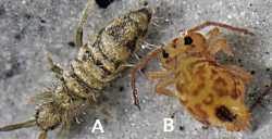
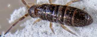
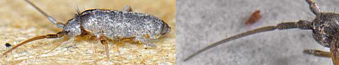
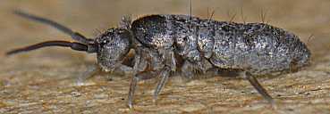
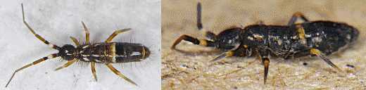
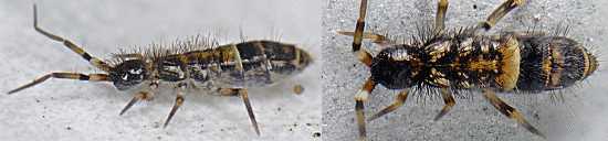
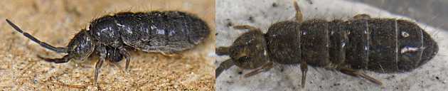
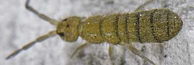
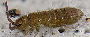
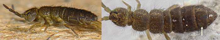
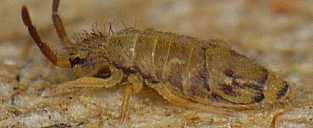
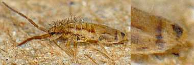
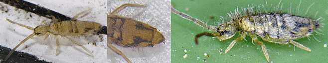
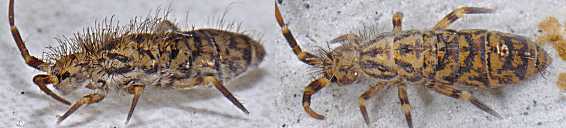
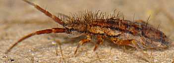
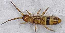
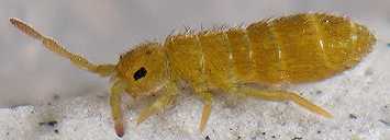
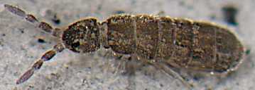
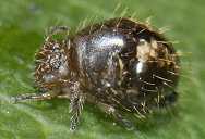
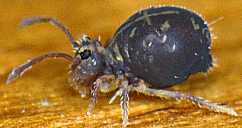
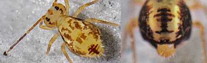
1(0). Body form elongate (fig.1a)................................ 2 Body form subglobular (fig.1b)............................ 23 |
|
 |
|
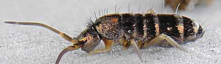
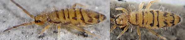
2(1). Body iridescent, covered with scales (fig.2)............... 3 Body not iridescent, not covered with scales................ 6 |
|
 |
|
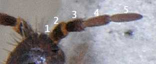
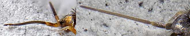
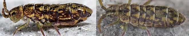
3(2). Apical antennomere about equal to subapical antennomere (fig.3); antenna with 5 segments (fig.4): basal antennomere subdivided.................... Heteromurus major Apical antennomere much shorter then subapical antennomere; antenna with 4 segments................................... 4 |
|
 |
|
 |
|
4(3). Third antennal segment subcylindric (fig.5)................ 5 Third antennal segment tapering (fig.6)...................... ............................... Pogonognathellus flavescens |
|
 |
|
 |
|
5(4). Body dark purplish with transversal goldish bands at intersegmental margins (fig.7).......... Tomocerus vulgaris Body uniform bluish-grey (fig.8)............. Tomocerus minor |
|
 |
|
 |
|
6(2). Head blackish............................................... 7 Head not blackish........................................... 8 |
|
7(6). Abdomen with distinct whitish transversal band (fig.9), (fig.10)............................ Orchesella cincta adult Abdomen without whitish transversal band (fig.11)............. .............................. Isotoma viridis var violacea |
|
 |
|
 |
|
 |
|
8(6). Body with long dorsal setae................................. 9 Body without long dorsal setae............................. 22 |
|
9(8). Body with few long dorsal setae............................ 10 Body with many long dorsal setae........................... 15 |
|
10(9). Body background colour not uniform......................... 11 Body background colour uniform............................. 13 |
|
11(10). Body pigmentation in longitudinal pattern.................. 12 Body pigmentation in transversal pattern (fig.12)............. .............................. Isotoma viridis var annulata |
|
 |
|
12(11). Lateral thoracic pigmentation distinctly patchy; middorsal segmental pigmentation in the shape of a crown (fig.13); tibiotarsi distinctly differently coloured than femora...... ...................................... Isotomurus maculatus Lateral thoracic pigmentation indistinct; middorsal segmental pigmentation in the shape of a solid line (fig.14); tibiotarsi and femora equally coloured. Isotomurus palustris |
|
 |
|
 |
|
13(10). Body background colour greenish (fig.15)....... Isotoma viridis Body background colour not greenish........................ 14 |
|
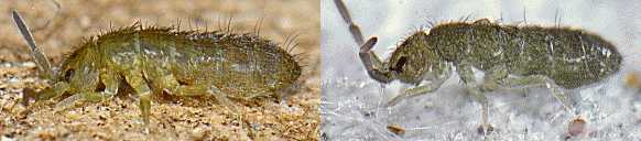 |
|
14(13). Body background colour brownish with pale random dot pattern (fig.16)......................... Isotoma anglicana juvenile Body background colour dark violet with distinct pale dot pattern lateral on abdomen (fig.17). Isotoma anglicana adult |
|
 |
|
 |
|
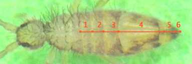
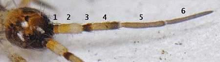
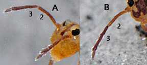
15(9). Antenna with 4 segments; trunk with one abdominal segment distinctly longer than the others (fig.18)............... 16 Antenna with 6 segments (fig.19): basal two antennomeres subdivided; trunk with subequal abdominal segments....... 20 |
|
 |
|
 |
|
16(15). Thoracic dorsal pigmentation absent........................ 17 Thoracic dorsal pigmentation present....................... 18 |
|
17(16). Dorsal pigmentation of the second and third abdominal segment in distinct transversal bands (fig.20)...................... ......................... Entomobrya nivalis forma dorsalis Dorsal pigmentation of the second and third abdominal segment in indistinct transversal patches (fig.18), (fig.21)........ ....................................... Entomobrya nicoleti |
|
 |
|
 |
|
18(16). Thoracic dorsal pigmentation in longitudinal bands (fig.22).... ....................................... Entomobrya muscorum Thoracic dorsal pigmentation in transversal bands.......... 19 |
|
 |
|
19(18). Dorsal abdominal pigmentation in irregular transversal bands (fig.23)........................... Entomobrya multifasciata Dorsal abdominal pigmentation in the shape of a capital U (fig.24).................................. Entomobrya nivalis |
|
 |
|
 |
|
20(15). Body pigmentation pattern in complex pattern of patches (fig.25).................................. Orchesella villosa Body pigmentation pattern in distinct bands................ 21 |
|
 |
|
21(20). Body pigmentation in longitudinal bands (fig.26)............... ..................................... Orchesella flavescens Body pigmentation in transversal bands (fig.27)............... ................................ Orchesella cincta juvenile |
|
 |
|
 |
|
22(8). Body colour greenish (fig.28)................ Desoria olivacea Body colour greyish (fig.29).................. Desoria tigrina |
|
 |
|
 |
|
23(1). Body background colour brownish............................ 24 Body background colour yellowish with patchy brown pigmentation............................................. 26 |
|
24(23). Body texture dull.......................................... 25 Body texture glossy (fig.30).................... Allacma fusca |
|
 |
|
25(24). Dorsum uniformly coloured (fig.31)............ Dicyrtoma fusca Dorsum with middorsal yellow longitudinal stripe (fig.32)..... ........................................ Dicyrtomina ornata 1 |
|
 |
|
 |
|
26(23). Dorsal posterior patch multi barred cross shaped (fig.33); second antennomere colour distinctly more pale then that of third antennomere, especially at the joint (fig.34b)........ ..................................... Dicyrtomina saundersi Dorsal posterior patch solid rectangular (fig.35); second antennomere colour gradually fading into that of third antennomere (fig.34a).................... Dicyrtomina ornata |
|
 |
|
 |
|
 |
|
Endnotes
1 Very dark colour form lacking the typical patchy pigmentation.
Allacma fusca
Body background colour brownish. Body form
subglobular (fig.1b). Body texture glossy (fig.30).
Desoria olivacea
Body not iridescent, not covered with scales.
Body without long dorsal setae. Body colour greenish (fig.28). Body form
elongate (fig.1a). Head not blackish.
Desoria tigrina
Body not iridescent, not covered with scales.
Body without long dorsal setae. Body colour greyish (fig.29). Body form
elongate (fig.1a). Head not blackish.
Dicyrtoma fusca
Body background colour brownish. Body form
subglobular (fig.1b). Body texture dull. Dorsum uniformly coloured (fig.31).
Dicyrtomina ornata
Body background colour yellowish with patchy
brown pigmentation. Body form subglobular (fig.1b). Dorsal posterior patch
solid rectangular (fig.35). Second antennomere colour gradually fading into
that of third antennomere (fig.34a).
Dicyrtomina ornata 1
Body background colour brownish. Body
form subglobular (fig.1b). Body texture dull. Dorsum with middorsal yellow
longitudinal stripe (fig.32).
Dicyrtomina saundersi
Body background colour yellowish with
patchy brown pigmentation. Body form subglobular (fig.1b). Dorsal posterior
patch multi barred cross shaped (fig.33). Second antennomere colour distinctly
more pale then that of third antennomere, especially at the joint (fig.34b).
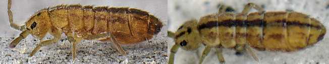
Entomobrya multifasciata
Antenna with 4 segments. Body not
iridescent, not covered with scales. Body with long dorsal setae. Body form
elongate (fig.1a). Body with many long dorsal setae. Dorsal abdominal
pigmentation in irregular transversal bands (fig.23). Head not blackish.
Thoracic dorsal pigmentation present. Thoracic dorsal pigmentation in
transversal bands. Trunk with one abdominal segment distinctly longer than the
others (fig.18).
Entomobrya muscorum
Antenna with 4 segments. Body not
iridescent, not covered with scales. Body with long dorsal setae. Body form
elongate (fig.1a). Body with many long dorsal setae. Head not blackish.
Thoracic dorsal pigmentation present. Thoracic dorsal pigmentation in
longitudinal bands (fig.22). Trunk with one abdominal segment distinctly longer
than the others (fig.18).
Entomobrya nicoleti
Antenna with 4 segments. Body not
iridescent, not covered with scales. Body with long dorsal setae. Body form
elongate (fig.1a). Body with many long dorsal setae. Dorsal pigmentation of
the second and third abdominal segment in indistinct transversal patches
(fig.18), (fig.21). Head not blackish. Thoracic dorsal pigmentation absent.
Trunk with one abdominal segment distinctly longer than the others (fig.18).
Entomobrya nivalis
Antenna with 4 segments. Body not iridescent,
not covered with scales. Body with long dorsal setae. Body form elongate
(fig.1a). Body with many long dorsal setae. Dorsal abdominal pigmentation in
the shape of a capital U (fig.24). Head not blackish. Thoracic dorsal
pigmentation present. Thoracic dorsal pigmentation in transversal bands. Trunk
with one abdominal segment distinctly longer than the others (fig.18).
Entomobrya nivalis forma dorsalis
Antenna with 4 segments. Body
not iridescent, not covered with scales. Body with long dorsal setae. Body
form elongate (fig.1a). Body with many long dorsal setae. Dorsal pigmentation
of the second and third abdominal segment in distinct transversal bands
(fig.20). Head not blackish. Thoracic dorsal pigmentation absent. Trunk with
one abdominal segment distinctly longer than the others (fig.18).
Heteromurus major
Antenna with 5 segments (fig.4): basal
antennomere subdivided. Apical antennomere about equal to subapical
antennomere (fig.3). Body iridescent, covered with scales (fig.2). Body form
elongate (fig.1a).
Isotoma anglicana adult
Body not iridescent, not covered with
scales. Body with long dorsal setae. Body background colour uniform. Body
background colour not greenish. Body background colour dark violet with
distinct pale dot pattern lateral on abdomen (fig.17). Body form elongate
(fig.1a). Body with few long dorsal setae. Head not blackish.
Isotoma anglicana juvenile
Body not iridescent, not covered with
scales. Body with long dorsal setae. Body background colour uniform. Body
background colour not greenish. Body background colour brownish with pale
random dot pattern (fig.16). Body form elongate (fig.1a). Body with few long
dorsal setae. Head not blackish.
Isotoma viridis
Body not iridescent, not covered with scales.
Body with long dorsal setae. Body background colour uniform. Body background
colour greenish (fig.15). Body form elongate (fig.1a). Body with few long
dorsal setae. Head not blackish.
Isotoma viridis var annulata
Body not iridescent, not covered
with scales. Body with long dorsal setae. Body background colour not uniform.
Body form elongate (fig.1a). Body pigmentation in transversal pattern
(fig.12). Body with few long dorsal setae. Head not blackish.
Isotoma viridis var violacea
Abdomen without whitish transversal
band (fig.11). Body not iridescent, not covered with scales. Body form
elongate (fig.1a). Head blackish.
Isotomurus maculatus
Body not iridescent, not covered with
scales. Body with long dorsal setae. Body background colour not uniform. Body
form elongate (fig.1a). Body pigmentation in longitudinal pattern. Body with
few long dorsal setae. Head not blackish. Lateral thoracic pigmentation
distinctly patchy. Middorsal segmental pigmentation in the shape of a crown
(fig.13). Tibiotarsi distinctly differently coloured than femora.
Isotomurus palustris
Body not iridescent, not covered with
scales. Body with long dorsal setae. Body background colour not uniform. Body
form elongate (fig.1a). Body pigmentation in longitudinal pattern. Body with
few long dorsal setae. Head not blackish. Lateral thoracic pigmentation
indistinct. Middorsal segmental pigmentation in the shape of a solid line
(fig.14). Tibiotarsi and femora equally coloured.
Orchesella cincta adult
Abdomen with distinct whitish
transversal band (fig.9), (fig.10). Body not iridescent, not covered with
scales. Body form elongate (fig.1a). Head blackish.
Orchesella cincta juvenile
Antenna with 6 segments (fig.19):
basal two antennomeres subdivided. Body not iridescent, not covered with
scales. Body with long dorsal setae. Body form elongate (fig.1a). Body
pigmentation in transversal bands (fig.27). Body pigmentation pattern in
distinct bands. Body with many long dorsal setae. Head not blackish. Trunk
with subequal abdominal segments.
Orchesella flavescens
Antenna with 6 segments (fig.19): basal two
antennomeres subdivided. Body not iridescent, not covered with scales. Body
with long dorsal setae. Body form elongate (fig.1a). Body pigmentation in
longitudinal bands (fig.26). Body pigmentation pattern in distinct bands. Body
with many long dorsal setae. Head not blackish. Trunk with subequal abdominal
segments.
Orchesella villosa
Antenna with 6 segments (fig.19): basal two
antennomeres subdivided. Body not iridescent, not covered with scales. Body
with long dorsal setae. Body form elongate (fig.1a). Body pigmentation
pattern in complex pattern of patches (fig.25). Body with many long dorsal
setae. Head not blackish. Trunk with subequal abdominal segments.
Pogonognathellus flavescens
Antenna with 4 segments. Apical
antennomere much shorter then subapical antennomere. Body iridescent, covered
with scales (fig.2). Body form elongate (fig.1a). Third antennal segment
tapering (fig.6).
Tomocerus minor
Antenna with 4 segments. Apical antennomere much
shorter then subapical antennomere. Body iridescent, covered with scales
(fig.2). Body uniform bluish-grey (fig.8). Body form elongate (fig.1a).
Third antennal segment subcylindric (fig.5).
Tomocerus vulgaris
Antenna with 4 segments. Apical antennomere
much shorter then subapical antennomere. Body iridescent, covered with scales
(fig.2). Body dark purplish with transversal goldish bands at intersegmental
margins (fig.7). Body form elongate (fig.1a). Third antennal segment
subcylindric (fig.5).