http://www.collembola.org/publicat/photogr.htm
-
Last updated on
2008.04.13
by Frans Janssens
Keith Brocklehurst,
114, Leicester Lane, Leamington Spa, Warwickshire, CV32 7HH, UK
Frans Janssens,
Department of Biology, University of Antwerp, Antwerp, B-2020, Belgium
The minute size of Collembola and their rapid unpredictable movements are
obvious problems in their live photography. Perhaps their illumination is also
a critical factor - cold fibre-optic light is best though expensive,
and not a complete answer by any means. Nevertheless, if they can be made to
remain still for a minute or so success will be more frequent. This is a
behavioural method of making them "sit" fairly still and all in one or more
chosen spots in a culture dish for a minute or more.
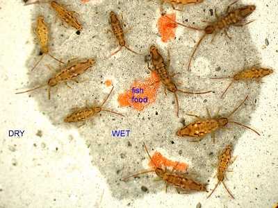 |
Fig.Ei. Entomobrya intermedia from the UK
Specimens drinking with eversed vesicles of collophore
2007 © Brocklehurst, K.
The images of Entomobrya intermedia (L = 2 mm) illustrate the spectacular and
rapid response they give after 72 hours dehydration in culture at comfortable
room temperature when a few drops of distilled water are quickly placed on
the dry culture medium. The DIY filler forming the base of the culture vessel
is whiter when dry and darker when wet; it contains tiny amounts of activated
carbon, which avoids a difficult white background. The live specimens were
collected by net and pooter from small garden plants. After dehydration five
tiny drops of water, spaced well apart in the culture, were added from a fine
pipette, and a few seconds later the springtails were all still and
distributed as in the photographs. Head movements when this species is eating
can be seen with magnification, but none was detected even from the occasional
individual which was near the fish food. So, my guess is that they were
applying their two-lobed ventral tube to the wet medium and absorbing water,
presumably through fine tubes. Further work should suggest a method !
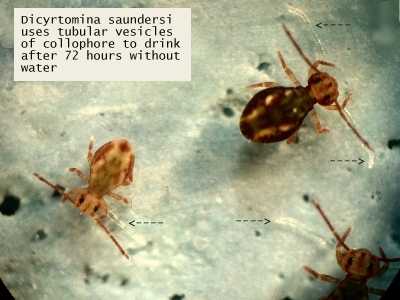 |
Fig.Ds. Dicyrtomina saundersi from the UK
Specimens drinking with eversed tubular vesicles of collophore
2007 © Brocklehurst, K.
The same behaviour occurs in Dicyrtomina saundersi, but here 2 vesicles
are suddenly extruded from the ventral tube and laid out flat on the moist
filler. The vesicles are fine and transparent with fine openings, and are
usually seen extended in preserved Dicyrtomids and sometimes tangled round
legs.
Taking photographs from the dorsal side by this method is straightforward
with modern digital cameras - it would be interesting to somehow get ventral
views. The images were made with a Nikon 995 camera.
Comments would be welcome.
 |
Fig.Do. Dicyrtomina ornata from France
Drinking with eversed tubular vesicles of collophore
2008.04.10 © Lebeaux, P.
This specimen of Dicyrtomina ornata from France presses its everted
tubular collophore vesicles against the wet substrate to 'drink'.
Acknowledgements
We would like to thank Philippe Lebeaux for his excellent contribution.
