 |
 |
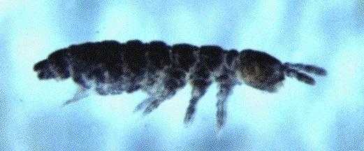
| Fig.1. Habitus lateral 40x Photograph Frans Janssens copyright © (1997) |
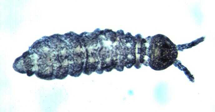
Fig.2. Habitus dorsal 32x Photograph Frans Janssens copyright © (1997) |
| Checklist of the Collembola: Aggregations of Hypogastrura ripperi Gisin, 1952 in Canada |
Note: This preliminary version of the document is . Results can change and conclusions can be rethought. It is recommended to not copy down, print or quote this text. To keep up to date, it is preferable to use a hyperlink in steadt.
This manuscript is a compilation of the cooperative communication between Dr Dan L. Johnson of the Agriculture and Agri-Food Canada Research Centre of Lethbridge, Canada and myself on the sudden appearance of small aggregations of a Hypogastrurid springtail in Lethbridge, Canada.
Vial 1 : 68 Collembola specimens in ethanol
Vial 2 : .. Collembola specimens in ethanol
Note : they were not 'fixed' : the transparant cuticula
came loose from the body, as if each specimen was wrapped
into a transparant plastic bag. Gisin's fixative 1
could have prevented this artefact.
Label :
Vial 1 :
Mar 26, 1997Vial 2 :
Lethbridge, AB, Canada
Dan Johnson
Hypogastruridae
Ceratophysella pseudarmata ?
Suspected Ceratophysella pseudarmata
D. Johnson, col
Mar 27-30, 1997
Lethbridge, Alberta, Canada
|
|
| Fig.1. Habitus lateral 40x Photograph Frans Janssens copyright © (1997) |
Fig.2. Habitus dorsal 32x Photograph Frans Janssens copyright © (1997) |
Based on Christiansen & Bellinger (1980), Ceratophysella cannot be confirmed : 'Nearctic Ceratophysella should have relatively large (never minute) anal spines'. Also Folsom's original description (1916) of Achorutes pseudarmatus specifies 'anal spines long'.
These preliminary observations indicate that it is not a Ceratophysella species.
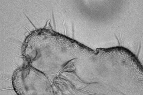 |
| Fig.3. Anal spine 100x Photograph Frans Janssens copyright © (1998) |
The key of the Norwegian Collembola of Fjellberg (1980) leads straightforward to Hypogastrura vernalis :
Poduridae
Hypogastrura
Empodium with small, narrow lamella.
Mucro with distinct tooth before apex.
1.2 mm. Dark bluish red. Anal spines short, stright. Often in large numbers in dry meadows close to sea.
Stach (1949) states about Neogastrura manubrialis : 'the limit between those segments [3rd and 4th antennal segments] sometimes indistinct'. What is certainly the case for this set of specimens. In fact, it is more or less valid for all Hypogastrura s.s. Also the fact that the antenna is thickest at the joint of the 3rd and 4th segment gives the impression (at lower magnification) that the 4th segment is fused with the 3rd. Stach (1949) notes on N. purpurescens dorso-lateral spotted lines. Those are clearly seen also in these specimens.
Tentative identification : Hypogastrura cf. vernalis (Carl, 1901)
Note that all characters apply to the blob specimens except that the specimens do not have knobbed tenent hairs... Therefore, the use of 'cf.'
After verifying Gisin's diagnosis of H.(H.) vernalis against some completely cleared specimens (gently heated on electrical heating plate in Gisin's clearing medium), the tentative identification has to be revised.
Following the key of the European Collembola of Gisin (1960) :
Hypogastrura (Hypogastrura)
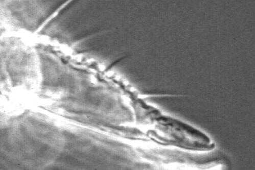 |
| Fig.4. Mucro with broad lamella 100x Photograph Frans Janssens copyright © (1998) |
- 1.5 mm. Grau-blau. Abd. V und VI dorsomedian mit stärkerer Hautkörnung. Ant. IV mit 3-4 äusseren und 2-3 inneren Riechhaaren. Klaue mit Innen-, ohne Seitenzahn. Tibiotarsus ohne Keulenhaar.
Ventraaltubus mit 4+4 Borsten (unveröff.). Tenaculum mit 4+4 Zähnen.
This identification is confirmed using the key of the Nearctic Collembola of Christiansen & Bellinger (1980). Both publications mention Neogastrura assimilis Stach 1949, nec Krausbauer 1898 as synomym of ripperi. Stach (1949) has a very detailed diagnosis of Neogastrura assimilis.
Final identification : Hypogastrura (Hypogastrura) ripperi Gisin, 1952
| Location : | Lethbridge, Albertha, Canada |
| Date : | 1997.03.26-30 |
| Leg. : | Dr Dan L. Johnson |
| Det. : | Frans Janssens |
| Col. : | Dr Dan L. Johnson |
Photographs of cleared specimens were made with a Fuji dia positive film of 100 ASA, type day-light. Dr De Bruyn of the RUCA, Antwerp, Belgium was so kind to provide for the scanned JPEG images.
At least the following photographs are to be made to document the identification :
| 95% ethanol | 750 ml |
| ethyl ether | 250 ml |
| glacial acetic acid | 30 ml |
| 40% formaldehyde | 3 ml |
| lactic acid | 100 ml |
| glycerol | 20 ml |
| 40% formaldehyde | 4 ml |